

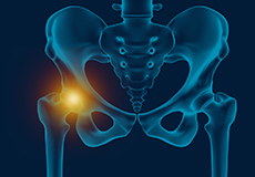
Hip
Hip Anatomy
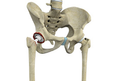
The hip joint is the largest weight-bearing joint in the human body. It is also referred to as a ball and socket joint and is surrounded by muscles, ligaments and tendons. The thighbone or femur and the pelvis join to form the hip joint.
Any injury or disease of the hip will adversely affect the joint's range of motion and ability to bear weight.
The hip joint is made up of the following:
- Bones and joints
- Ligaments of the joint capsule
- Muscles and tendons
- Nerves and blood vessels that supply the bones and muscles of the hip
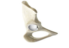
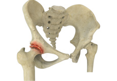
Bones and Joints
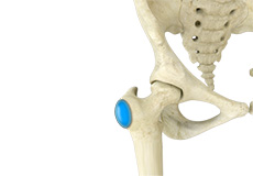
The hip joint is the junction where the hip joins the leg to the trunk of the body. It is comprised of two bones: the thighbone or femur, and the pelvis, which is made up of three bones called ilium, ischium and pubis.
The ball of the hip joint is made by the femoral head while the socket is formed by the acetabulum. The acetabulum is a deep, circular socket formed on the outer edge of the pelvis by the union of three bones: ilium, ischium and pubis. The lower part of the ilium is attached by the pubis while the ischium is considerably behind the pubis. The stability of the hip is provided by the joint capsule or acetabulum and the muscles and ligaments that surround and support the hip joint.
The head of the femur rotates and glides within the acetabulum. A fibrocartilaginous lining called the labrum is attached to the acetabulum and further increases the depth of the socket.
The femur is one of the longest bones in the human body. The upper part of the thighbone consists of the femoral head, femoral neck, and greater and lesser trochanters. The head of the femur joins the pelvis (acetabulum) to form the hip joint. Next to the femoral neck, there are two protrusions known as greater and lesser trochanters which serve as sites of muscle attachment.
Articular cartilage is the thin, tough, flexible and slippery surface lubricated by synovial fluid that covers the weight-bearing bones of the body. It enables smooth movements of the bones and reduces friction.
Ligaments
Ligaments are fibrous structures that connect bones to other bones. The hip joint is encircled with ligaments to provide stability to the hip by forming a dense and fibrous structure around the joint capsule. The ligaments adjoining the hip joint include:
- Iliofemoral ligament: This is a Y-shaped ligament that connects the pelvis to the femoral head at the front of the joint. It helps in limiting over-extension of the hip.
- Pubofemoral ligament: This is a triangular shaped ligament that extends between the upper portion of the pubis and the iliofemoral ligament. It attaches the pubis to the femoral head.
- Ischiofemoral ligament: This is a group of strong fibers that arise from the ischium behind the acetabulum and merge with the fibers of the joint capsule.
- Ligamentum teres: This is a small ligament that extends from the tip of the femoral head to the acetabulum. Although it has no role in hip movement, it does have a small artery within that supplies blood to a part of the femoral head.
- Acetabular labrum: The labrum is a fibrous cartilage ring which lines the acetabular socket. It deepens the cavity increasing the stability and strength of the hip joint.
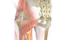
Muscles and Tendons
A long tendon called the iliotibial band runs along the femur from the hip to the knee and serves as an attachment site for several hip muscles including the following:
- Gluteal: These are the muscles that form the buttocks. There are three muscles (gluteus minimus, gluteus maximus, and gluteus medius) that attach to the back of the pelvis and insert into the greater trochanter of the femur.
- Adductors: These muscles are in the thighs which help in adduction, the action of pulling the leg back towards the midline.
- Iliopsoas: This muscle is in front of the hip joint and provides flexion. It is a deep muscle that originates from the lower back and pelvis, and extends up to the inside surface of the upper part of the femur.
- Rectus femoris: This is the largest band of muscles located in front of the thigh. They are also called hip flexors.
- Hamstring muscles: These begin at the bottom of the pelvis and run down the back of the thigh. Because they cross the back of the hip joint, they help in extension of the hip by pulling it backwards.
Nerves and Arteries
Nerves of the hip transfer signals from the brain to the muscles to aid in hip movement. They also carry the sensory signals such as touch, pain, and temperature back to the brain.
The main nerves in the hip region include the femoral nerve in the front of the femur and the sciatic nerve at the back. The hip is also supplied by a smaller nerve known as the obturator nerve.
In addition to these nerves, there are blood vessels that supply blood to the lower limbs. The femoral artery, one of the largest arteries in the body, arises deep in the pelvis and can be felt in front of the upper thigh.
Hip Movements
All the anatomical parts of the hip work together to enable various movements. Hip movements include flexion, extension, abduction, adduction, circumduction, and hip rotation.
Hip Bursitis
Hip bursitis is a painful condition caused by the inflammation of a bursa in the hip. Bursae are fluid-filled sacs present in the joints between bone and soft tissue to reduce friction and provide cushioning during movement.
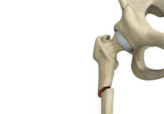
Hip Fracture
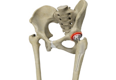
A hip fracture is a break that occurs near the hip in the upper part of the femur or thighbone. The thighbone has two bony processes on the upper part - the greater and lesser trochanters. The lesser trochanter projects from the base of the femoral neck on the back of the thighbone.
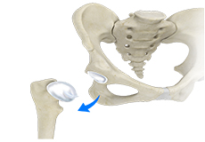
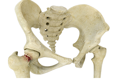
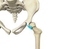
Hip Dislocation
Hip dislocation occurs when the head of the femur moves out of the socket. The femoral head can dislocate either backward (posterior dislocation) or forward (anterior dislocation).
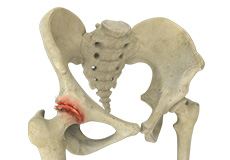
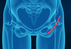
Hip Labral Tear
A hip labral tear is an injury to the labrum, the cartilage that surrounds the outside rim of your hip joint socket.
Subtrochanteric Hip Fracture
A hip fracture is a break that occurs near the hip in the upper part of the femur or thighbone. The thighbone has two bony processes on the upper part - the greater and lesser trochanters. The lesser trochanter projects from the base of the femoral neck on the back of the thighbone.
Developmental Dysplasia
Developmental dysplasia of the hip (DDH) or hip dysplasia is a condition that is seen in infants and young children because of developmental problems in the hip joint. The femur (thighbone) partially or completely slips out of the hip socket leading to dislocation at the hip joint.
Osteoarthritis of the Hip
Osteoarthritis, also called degenerative joint disease, is the most common form of arthritis. It occurs most often in the elderly. This disease affects the tissue covering the ends of bones in a joint called cartilage.
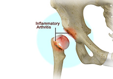
Inflammatory Arthritis of the Hip
The inflammation of the joints is referred to as arthritis. Inflammation arises when the smooth lining called cartilage at the ends of bones wears away. In some cases, the inflammation is caused when the lining of the joint becomes inflamed as part of an underlying systemic disease.
Periprosthetic Hip Infection
A very small percentage of patients (less than 1%) who undergo hip replacement may develop an infection around the hip joint following surgery. This infection is called a periprosthetic hip infection.
Anterior Hip Replacement
Anterior hip replacement surgery is performed under general anesthesia or regional anesthesia. You will lie down on your back, on a special operating table that enables your surgeon to perform the surgery from the front of the hip.
Hip Fracture ORIF
A hip fracture is a break that occurs near the hip in the upper part of the femur or thighbone. The thighbone has two bony processes on the upper part - the greater and lesser trochanters. The lesser trochanter projects from the base of the femoral neck on the back of the thighbone.
Correction of a Failed Hip Replacement
Reoperation of a total hip replacement to resolve a painful hip condition arising out of a damaged or worn out prosthesis (artificial hip joint) is known as correction of a failed hip replacement.
Correction of a Painful Hip Replacement
Reoperation of a total hip replacement to resolve a painful hip condition arising out of a damaged or worn out prosthesis (artificial hip joint) is known as correction of a painful hip replacement.
Correction of a Loose Hip Replacement
Reoperation of a total hip replacement to resolve a painful hip condition and loss of motion due to a loosened prosthesis (artificial hip joint) is known as correction of a loose hip replacement. This loosening occurs due to wear and tear of the implant surfaces and subsequent weakening of the surrounding bone.
Hip Fracture Surgery
Hip fractures involve a break that occurs near the hip in the upper part of the femur or thigh bone. The thigh bone has two bony processes on the upper part - the greater and lesser trochanters. The lesser trochanter projects from the base of the femoral neck on the back of the thigh bone.
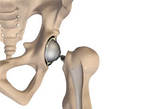
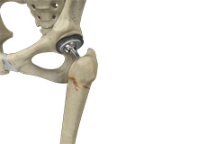
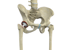
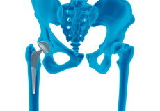
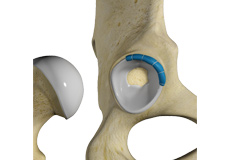
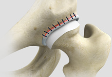
Total Hip Replacement
Total hip replacement is a surgical procedure in which the damaged cartilage and bone is removed from the hip joint and replaced with artificial components.
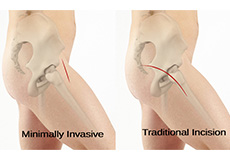
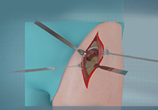
Minimally Invasive Total Hip Replacement
Traditionally, total hip replacement will be performed through a 10–12-inch-long incision made on the side of your hip. A minimally invasive approach has been developed in recent years where surgery is performed through one or two smaller incisions rather than the single long incision as in the traditional approach.
Revision Hip Replacement
During total hip replacement, the damaged cartilage and bone are removed from the hip joint and replaced with artificial components. At times, hip replacement implants can wear out for various reasons and may need to be replaced with the help of a surgical procedure known as revision hip replacement surgery.
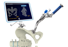
Computer-assisted Hip Replacement
Computer-assisted hip replacement is an image-guided, minimally invasive surgical procedure to replace your diseased or damaged hip with an artificial device using the assistance of computer software.
Hip Trauma Reconstruction
Hip trauma is an injury in the hip due to the impact caused by incidents such as a car accident or a hard fall. The injury can be a bone break or dislocation or both.
Hip Reconstruction
Hip reconstruction is a surgery to repair or replace a damaged hip joint that causes pain and limits your movement.
Outpatient and Rapid Recovery Hip Replacement
Outpatient joint replacement is a surgery that does not require an overnight hospital stay. Joint replacement is a surgical procedure in which the worn, damaged surfaces of the joint are removed and replaced with new artificial parts.
Complex Hip Replacement
Coming soon

Activities After Hip Replacement
Hip replacement is a surgery performed to replace parts of a diseased hip joint with a prosthesis. The goal of hip replacement is to eliminate pain and enable you to return to your normal activities. You can help in the recovery and improve the outcomes of the procedure by following certain precautions and changing the way you carry out your daily activities.
Comprehensive Care of Hip Osteoarthritis
Osteoarthritis, also called degenerative joint disease, is the most common form of arthritis. It occurs most often in the elderly. This disease affects the tissue covering the ends of bones in a joint called cartilage.